
Page 33 - Application Handbook - Liquid Chromatography
P. 33
LAAN-A-LC-E245
Application High Performance Liquid Chromatography
News Simultaneous Determination of Polycyclic Aromatic
Hydrocarbons Using the Prominence-i Integrated
No.L468 High Performance Liquid Chromatograph
Many polycyclic aromatic hydrocarbons exhibit Table 2 Analytical Conditions
fluorescence, and can therefore be detected with high
selectivity and high sensitivity using a fluorescence Detector : UV at 230 nm
RF-20Axs
detector. Previously, in Application News No. 393 and 0.0 - 10.0 min Ex. at 270 nm, Em. at 330 nm, Gain : X1
No. 441A, we introduced examples of the simultaneous 10.0 - 12.8 min Ex. at 250 nm, Em. at 370 nm, Gain : X1
analysis of polycyclic aromatic hydrocarbons (PAHs) 12.8 - 16.0 min Ex. at 330 nm, Em. at 430 nm, Gain : X4
using a fluorescence detector. However, of the sixteen 16.0 - 21.0 min Ex. at 270 nm, Em. at 390 nm, Gain : X1
21.0 - 27.6 min Ex. at 290 nm, Em. at 430 nm, Gain : X1
polycyclic aromatic hydrocarbons designated as 27.6 - 30.0 min Ex. at 370 nm, Em. at 460 nm, Gain : X16
"priority pollutants" by the U.S. Environmental 30.0 - 40.0 min Ex. at 270 nm, Em. at 330 nm, Gain : X1
Protection Agency (EPA), acenaphthylene alone does Cell Temp. : 28 °C
not exhibit fluorescence. Therefore, a single
fluorescence detector cannot be used for simultaneous
analysis of all sixteen of these PAHs. However, the
Prominence-i, which incorporates a UV detector, can be (a)
connected to the RF-20Axs fluorescence detector as an 3.00 mV
optional detector, permitting simultaneous analysis of 2.75 LC-2030 UV at 254 nm
all sixteen polycyclic aromatic hydrocarbons. Here, using 2.50
two analytical methods, one with the wavelength 2.25
switching mode and the other using simultaneous 2.00
measurement at multiple wavelengths, we introduce an 1.75 6
example of simultaneous analysis of the 16 PAHs. 1.50 11
1.25 2
n Analysis of Polycyclic Aromatic Hydrocarbons 1.00 1 5 10 13 16
Using Wavelength Switching Mode 0.75 4 7 9 12 14 15
0.50
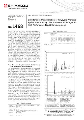
Fig. 1 (a) shows the chromatogram obtained using a UV 3 8
detector for analysis (detection wavelength: 254 nm), 0.25
and Table 1 shows the analytical conditions that were 0.00
used. Although good separation was obtained using -0.25 0.0 5.0 10.0 15.0 20.0 25.0 30.0 min
these conditions, the insufficient sensitivity obtained
with UV absorption alone is due to the low absorption (b) mV (×1000)
of ultraviolet light by the PAHs. LC-2030 UV and RF-20Axs
Fig. 1 (b) shows chromatograms obtained via 1.25
simultaneous analysis of sixteen polycyclic aromatic 3
hydrocarbons using the wavelength switching mode 1.00
according to component group, accomplished by
connecting the Prominence-i to an RF-20Axs detector. 0.75
Table 2 shows the analytical conditions used for 1
conducting analysis via combination of the fluorescence 0.50 4 12 15
detector and UV detector. The non-fluorescent 2 9 13
acenaphthylene is detected by the UV absorption 0.25 8 10 11 14 230 nm (UV)
detector at 230 nm, in the vicinity of the wavelength at 5 6 7 16 RF-20Axs
which it exhibits the maximum absorption. Using these 0.00
analytical conditions, good sensitivity and separation of 0.0 5.0 10.0 15.0 20.0 25.0 30.0 min
the sixteen components is achieved, permitting analysis 1. Naphthalene (1.0 mg/L) 2. Acenaphthylene (2.0 mg/L)
of each component at the optimum wavelength. 3. Acenaphthene (1.0 mg/L) 4. Fluorene (0.2 mg/L)
5. Phenanthrene (0.1 mg/L) 6. Anthracene (0.1 mg/L)
7. Fluoranthene (0.2 mg/L) 8. Pyrene (0.1 mg/L)
Table 1 Analytical Conditions 9. Benzo[a]anthracene (0.1 mg/L) 10. Chrysene (0.1 mg/L)
11. Benzo[b]fluoranthene (0.2 mg/L) 12. Benzo[k]fluoranthene (0.1 mg/L)
Column : RESTEK Pinacle Ⅱ PAH (250 mm L. × 4.6 mm I.D., 4 μm) 13. Benzo[a]pyrene (0.1 mg/L) 14. Dibenzo[a, h]anthracene (0.2 mg/L)
Mobile Phase : A: Water 15. Benzo[g, h, i]perylene (0.2 mg/L) 16. Indeno[1, 2, 3-cd]pyrene (0.1 mg/L)
B: Acetonitrile
Column Temp. : 40 °C
Time Program : B Conc. 60 % (0 - 5 min) → 100 % (30 - 35 min) Fig. 1 (a) UV Chromatogram of Standard Mixture of 16 PAHs
→ 60 % (35 - 40 min) (b) Chromatogram of Standard Mixture of 16 PAHs
Flowrate : 1.5 mL/min Analyzed by the RF-20Axs and UV Detector
Injection : 5 μL Note: The peak intensity of the chromatogram of Fig. 1 (b) is
Detector : UV at 254 nm displayed as 10 times that of the actual chromatogram.