Page 14 - Shimadzu EPMA-8050G
P. 14

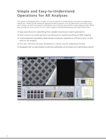
Simple and Easy-to-Understand                                                                                      Advanced image processing technology


                     Operations for All Analyses                                                                                        brings out even High Performance


                     This system is equipped with a number of new functions to enable simple and easy-to-understand                       Jag-Reduction Function (Standard Equipment)
                     operations. These include advanced operational performance, with all operations controlled using
                     just a mouse, as well as an easy-to-understand user interface and a built-in easy analysis mode. While             Reduces the whisker-like distortion (caused by mechanical vibration or magnetic  eld  uctuations) in SEM
                     easy enough for even novices to use, it also supports sophisticated analysis by experienced users.                 images that becomes more noticeable at higher magni cation. It uses state-of-the-art image processing
                                                                                                                                        technology that relocates each pixel to the optimum position and reconstructs the image. It reduces image
                     ˔ Easy operations for everything from sample mounting to report generation                                         distortion without compromising the original resolution, resulting in a sharper SEM image.
                     ˔ Even novices can easily perform everything from sample searching to SEM imaging
                     ˔ Unprecedented operability dramatically heightens operational efficiency prior to the
                        start of the analysis
                     ˔ The user interface has been designed in a visual, easy-to-understand format

                     ˔ Equipped with an easy analysis mode that automates all processes up to generating reports


                                                                                                                                                          Before Correction                      After Correction (Jag-Reduction)
                                                                                                                                        The above image is an example of a SEM image of a gold-deposited particle taken at a magni cation of
                                                                                                                                        ×100,000 and compared before and after correction. The corrected image not only reduces the edge
                                                                                                                                        distortion of the large particles, but also clearly shows the shape of the small particles.


                                                                                                                                          View Hold Function (Standard Equipment)

                                                                                                                                        This function prevents the electron beam from deviating from the target foreign matter and outputting the
                                                                                                                                        composition information of the wrong part when performing qualitative analysis of minute foreign matter.


                                                                                                                                        This is an example of the screen with the View Hold function activated. The electron beam is controlled so
                                                                                                                                        that the target in the middle of the SEM image does not move within the rectangular area.
                                                                                                                                                                                                                        EPMA-8050G
                                                                                                                                                                                                                         Electron Probe Microanalyzer
        14                                                                                                                                                                                                                                  15
   9   10   11   12   13   14   15   16   17   18   19