Page 26 - Pharmaceutical- Guide to Biopharmaceutical
P. 26

Characterization                Quality Control


                     Easily Determine Protein Secondary Structures                                                                      IRTracer-100


                               Analysis of Protein Secondary Structures                                                                                    benefits

                               —Analysis on Changes of Secondary Structures in Egg White Proteins Caused by Thermal Denaturation—                                                                                                            Cell Line Optimization
                                                                                                 click here                                     •   By using a heatable three-reflection ATR accessory, infrared spectra can be obtained
                                                                                                                                                  from proteins in a heated environment.
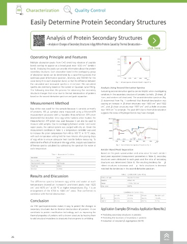
                     Operating Principle and Features                             40℃
                                                                              0.06  50℃                                                         •   Slight variations in infrared spectral shapes can be shown clearly by calculating the
                                                                                  60℃
                                                                                  70℃
                     Multiple absorption peaks from C=O stretching vibration of peptide   Abs  80℃
                                                                                  90℃
                                                                                  100℃                                                            second derivative of infrared spectra obtained.
                     bonds overlap to appear as a broad peak near 1650 cm  (amide I   0.04
                                                         -1
                     band). Analyzing the peaks can provide information about the protein                                                       •   The secondary structures of proteins can be analyzed by separating amide I band          Culture
                                                                              0.02
                     secondary structures. Each absorption band in the overlapping group                                                          peaks in second-derivative spectra.
                     of absorption bands can be determined by a curve-fitting process that
                                                                              0.00
                     optimizes peak information (position, intensity, and FWHM) for the   1700  1675  1650  1625  1600
                                                                                                 cm -1
                     curve being fit to each absorption band, so that the difference between   Fig. 1   Infrared Spectra of Amide I Band in Egg White
                     the calculated and measured spectra is minimized. The calculated
                     spectra are commonly based on the Lorenz or Gaussian curve fitting.   Analysis Using Second-Derivative Spectra
                     The following describes the process for observing the secondary   Evaluating second-derivative spectra can be helpful when investigating
                     structural changes that occur due to thermal denaturation of proteins   variations in the secondary structure of proteins (α-helix, β-sheet, β
                     based on the second-derivative spectrum and peak separation.  -turn, and random coil structures). The second-derivative spectrum (Fig.
                                                                   2) determined from Fig. 1 confirmed that thermal denaturation was                                                                                                         Purification
                     Measurement Method                            causing an increase in β-sheet structures near 1693 cm  and 1622
                                                                                                      -1
                                                                   cm , and β-sheet structures near 1637 cm  and α-helix structures
                                                                                              -1
                                                                     -1
                     Egg white was used for the sample because it consists primarily   near 1655 cm  to untangle. The peak shift due to thermal denaturation
                                                                           -1
                     of proteins. 60 μL  samples  were measured using a MicromATR   suggests the status of hydrogen bonds may have changed.
                     measurement accessory with a heatable three-reflection ATR prism
                     (diamond/ZnSe) installed. Since egg white hardens when heated, the
                     three-reflection ATR prism was used because it can also be used to
                     measure solid samples. Due to overlapping between amide I and water
                     vapor peaks, the optical system was purged with dry air. Given the     Decreases
                                                                                        Decreases
                     measurement conditions in Table 1, a temperature controller was used
                     to increase the prism temperature from 40 to 100 °C in 10 °C steps,   Increases                                                                                                                                         Characterization
                     with each temperature setting held for two minutes after placing drops   Random   Increases
                     of egg white to ensure adequate heat transfer before measuring. To   β -Sheet  β -Turn  α-Helix  Coil   β -Sheet
                     eliminate the effects of moisture in the egg white, analysis was based on
                     difference spectra calculated by subtracting the spectrum for water at   Fig. 2   Second-Derivative Spectra of Spectra in Fig. 1
                     each temperature.                             Amide I Band Peak Separation                                         Specifications
                                                                   Based on the peak wavenumber and area value for each amide I
                                   Table 1   Measurement Conditions                                                                      Instrument            IRTracer-100
                                                                   band peak separated (measurement parameters in Table 2), secondary
                     Resolution       4 cm -1
                                                                   structures were attributed to each peak and the ratio of secondary    Interferometer        Michelson interferometer (30° incident angle)
                     Accumulation     100
                     Apodization function  Sqr-Triangle            structures was determined (Table 3). The resulting tendency for   β                         Equipped with Advanced Dynamic Alignment system                               Quality Control
                     Zero filling     4 times                      -sheet structures to increase and   α -helix structures to decrease                         Sealed interferometer with Automatic Dehumidifier
                     Detector         DLATGS                       matched the tendencies in the second-derivative spectrum.             Optical system        Single-beam optics
                                                                                                                                         Beam splitter         Germanium-coated KBr for Middle IR (Standard)
                                                                                Table 2   Conditions for Curve Fitting
                     Results and Discussion                                                                                                                    Germanium-coated CsI for Middle/Far IR (Optional)
                                                                   Peak curve type
                                                                                    Gaussian function
                                                                                                                                                               Silicon-coated CaF2 for Near IR (Optional)
                                                                   Baseline         Offset 1 Pt
                     The difference spectra  between  egg white and  water  at  each
                                                                   Range            1710 to 1580 cm -1                                   Light source          High-energy ceramic for Middle/Far IR (Standard) with 3 years guaranteed
                     temperature showed an increase in prominent peaks near 1625   Max. error  0.01%                                                           Tungsten lamp for Near IR (Optional)
                                 -1
                       -1
                     cm  and 1675 cm  at 60 °C or higher temperatures (Fig. 1 is an
                                              -1
                     enlargement of the 1700 to 1600 cm  area). That confirmed its                                                       Detector              DLATGS detector with temperature control for Middle/Far IR (Standard)
                                                                                  Conditions for Curve Fitting                                                 MCT (Hg–Cd–Te) with liquid nitrogen cooling for Middle/Near IR (Optional)     Pharmacokinetics
                     correlation with thermal denaturation.
                                                                            α-helix  β-sheet  β-turn  Random coil                                              InGaAs for Near IR (Optional)
                                                                   40 °C    30.3 %  37.9 %   16.4 %   15.4 %                                                              -1
                     Conclusion                                    100 °C   15.1 %  47.6 %   29.7 %   7.7 %                              Wavenumber range      7,800 to 350 cm  (Standard)
                                                                                                                                                                           -1
                                                                                                                                                               12,500 to 240 cm  (Optional)
                                                                                                                                                                                 -1
                     An FTIR spectrophotometer makes it easy to predict the changes in                                                   Resolution            0.25, 0.5, 1, 2, 4, 8, 16 cm  (Middle/Far IR)
                                                                                                                                                                         -1
                     secondary structures due to thermal denaturation of proteins. It can   Application Examples (Shimadzu Application News No.)               2, 4, 8, 16 cm  (Near IR)
                     contribute to protein modification technology, such as improving the                                                Dimensions            W 600 mm × D 665 mm × H 295 mm
                     thermal properties of proteins with a known structure by heating them   • Predicting secondary structures in proteins                                                                                                   Others
                     to add structural mutations to structures that are prone to unfolding.  • Predicting the locations of mutations in proteins  Weight       47 kg
                                                                   • Evaluation of amyloid-β aggregation (A619)
                                                                                                                                         Measurements          Spectrum measurement, continuous measurement, atmospheric correction measurement, continuous
                                                                                                                                                               measurement using ASC, simple measurement mode
        26                                                                                                                                                                                                                                  27
                                                                                                            index                                                                                                                  index
   21   22   23   24   25   26   27   28   29   30   31