
Page 26 - EDX-7200
P. 26
Small Spot Analysis Kit (Option)
For Analysis of Small Contaminants and Defect Analysis in Small Regions
This option can be used to analyze even smaller areas by replacing the collimator plate and sample
observation camera. It is especially useful for analyzing trace foreign matter and defects in micro areas,
and measuring plating thickness.
Minimum 0.3 mm X-Ray Irradiation Diameter
The excitation X-rays can be collimated to 0.3 mm in diameter, which is effective for the
high-accuracy analysis of small contaminants and for defect analysis in small regions,
Sample Image at an Irradiation Diameter
analyses difficult with standard specifications (minimum 1 mm in diameter). of 0.3 mm (Extended Zoom)
Sample: stainless powder (approx. 0.1 mm)
Enlarged Sample Images without Image Quality Degradation collected on filter paper
This system supports smaller samples, which heightens the visibility of sample
observation images. Users can switch to an enlarged image approximately 2.5 times
Irradiation area
larger than a previous image, without image quality degradation. 0.3 mm in diameter
(yellow circle)
Automatic Four-Stage Switching Between 0.3, 1, 3, and 10 mm in Diameter Irradiation area
1 mm in diameter
The irradiation diameter automatically switches between 0.3, 1, 3, and 10 mm in diameter. (green circle)
This system supports not only the analysis of small spots but also macro composition
analysis at 10 mm in diameter.
Note: The irradiation diameter is the size on the sample surface. Metal Plated Terminals
(At 1 mm in diameter, the irradiation area is not
Irradiation diameter Extended zoom button within the measurement area, so measurements
switching buttons (Enlargement to approx.
2.5 times the camera image) are impossible. At 0.3 mm in diameter,
measurements are possible.)
PCEDX Navi Sample Image Setting Fields
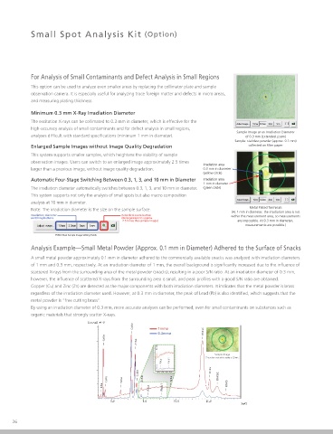
Analysis Example—Small Metal Powder (Approx. 0.1 mm in Diameter) Adhered to the Surface of Snacks
A small metal powder approximately 0.1 mm in diameter adhered to the commercially available snacks was analyzed with irradiation diameters
of 1 mm and 0.3 mm, respectively. At an irradiation diameter of 1 mm, the overall background is significantly increased due to the influence of
scattered X-rays from the surrounding area of the metal powder (snacks), resulting in a poor S/N ratio. At an irradiation diameter of 0.3 mm,
however, the influence of scattered X-rays from the surrounding area is small, and peak profiles with a good S/N ratio are obtained.
Copper (Cu) and Zinc (Zn) are detected as the major components with both irradiation diameters. It indicates that the metal powder is brass
regardless of the irradiation diameter used. However, at 0.3 mm in diameter, the peak of Lead (Pb) is also identified, which suggests that the
metal powder is “free cutting brass”.
By using an irradiation diameter of 0.3 mm, more accurate analyses can be performed, even for small contaminants on substances such as
organic materials that strongly scatter X-rays.
CuKa 1mmø
0.3mmø RhKaC
CaKa
ZnKa
ZnKb
Sample Image
(The yellow circle at the center is 0.3 mm.)
PbLa PbLb1
CuKb RhKa
CaKb FeKa ZnKb SrKa RhKbC
KKa PbLa PbLb1 RhKb
EDX-7200
26 Energy Dispersive X-Ray Fluorescence Spectrometer