
Page 17 - LifeScience Solution for FNIRS
P. 17
For Research Use Only. Not for use in diagnostic procedures.
Investigating Inner Speech Light to Measure Brain Function Principle of Using Near Infrared
Data Imaging Optical Brain-Function
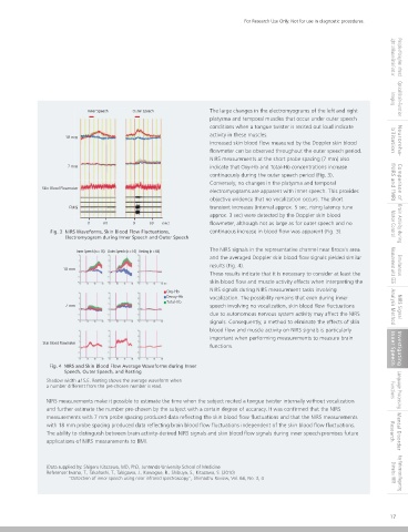
NIRS signals from the head surface were used to estimate the time when the subject made inner speech without vocalization and to Inner Speech Outer Speech The large changes in the electromyograms of the left and right
guess a number between 1 and 5 reechoes by the subject in his/her mind. platysma and temporal muscles that occur under outer speech
Two separations between transmitter and receiver optical fiber probes (7 and 18 mm spacing), as shown in Fig. 1, were used to conditions when a tongue twister is recited out loud indicate
measure the NIRS signals across the entire left and right forehead. Simultaneous measurements of the scalp blood flow and 18 mm activity in these muscles.
electromyograms were performed to evaluate the effects of skin blood flow and muscle blood flow on NIRS signals. Increased skin blood flow measured by the Doppler skin blood bilitation Neuroreha-
flowmeter can be observed throughout the outer speech period.
NIRS measurements at the short probe spacing (7 mm) also
Photo-transmitter probe
7 mm indicate that Oxy-Hb and Total-Hb concentrations increase
Photo-receiver probe continuously during the outer speech period (Fig. 3).
Conversely, no changes in the platysma and temporal fNIRS and fMRI Comparison of
Measurement channels Skin Blood Flowmeter
electromyograms are apparent with inner speech. This provides
Doppler
objective evidence that no vocalization occurs. The short
Platysma EMG EMG transient increases (interval approx. 5 sec, rising latency tune
approx. 3 sec) were detected by the Doppler skin blood
Temporal EMG
0 60 0 60 (sec) flowmeter, although not as large as for outer speech and no Motor Control Brain Activity during
Fig. 3 NIRS Waveforms, Skin Blood Flow Fluctuations, continuous increase in blood flow was apparent (Fig. 3).
Electromyogram during Inner Speech and Outer Speech
Inner Speech (n = 16) Outer Speech (n = 16) Resting (n = 64) The NIRS signals in the representative channel near Broca's area
and the averaged Doppler skin blood flow signals yielded similar
results (Fig. 4). Measurement with EEG Simultaneous
18 mm
These results indicate that it is necessary to consider at least the
skin blood flow and muscle activity effects when interpreting the
Oxy-Hb NIRS signals during NIRS measurement tasks involving
Deoxy-Hb vocalization. The possibility remains that even during inner
Fig. 1 Multiresolution Analysis Total-Hb
7 mm speech involving no vocalization, skin blood flow fluctuations Analysis Method NIRS Signal
due to autonomous nervous system activity may affect the NIRS
The probe channel positions are projected onto the MRI brain surface image. Probes are placed at 18 mm spacing in a 3×5
arrangement over the entire left and right forehead (44 ch), and at 7 mm spacing on the left forehead (1 ch). signals. Consequently, a method to eliminate the effects of skin
blood flow and muscle activity on NIRS signals is particularly
Fig. 2 shows typical NIRS signals when the subject recites a tongue twister internally without vocalization when the chosen number is important when performing measurements to measure brain
read. Multiple channels containing Broca's area (representative channel 11) show a transient increase in Oxy-Hb and Total-Hb Skin Blood Flowmeter functions.
concentrations when the chosen number 3 is read. Inner Speech Investigating
Fig. 4 NIRS and Skin Blood Flow Average Waveforms during Inner
Speech, Outer Speech, and Resting
Shadow width ±1S.E. Resting shows the average waveform when
a number different from the pre-chosen number is read. Functions Language Processing
NIRS measurements make it possible to estimate the time when the subject recited a tongue twister internally without vocalization
and further estimate the number pre-chosen by the subject with a certain degree of accuracy. It was confirmed that the NIRS
measurements with 7 mm probe spacing produced data reflecting the skin blood flow fluctuations and that the NIRS measurements
with 18 mm probe spacing produced data reflecting brain blood flow fluctuations independent of the skin blood flow fluctuations.
The ability to distinguish between brain activity-derived NIRS signals and skin blood flow signals during inner speech promises future Research Mental Disorder
applications of NIRS measurements to BMI.
Fig. 2 Typical NIRS Waveforms Blood Flow Fluctuations in Broca's Area (representative channel 11).
(Data supplied by: Shigeru Kitazawa, MD, PhD, Juntendo University School of Medicine
NIRS waveform results from four subjects were visually evaluated by five researchers. They were able to determine the number chosen Reference: Iwano, T., Takahashi, T., Takigawa, J., Kawagoe, R., Shibuya, S., Kitazawa, S. (2010) Shimadzu fNIRS Key References Regarding
"Detection of inner speech using near infrared spectroscopy", Shimadzu Review, Vol. 66, No. 3, 4
by the subject with 73 % accuracy.
16 17