
Page 18 - Shimadzu Journal vol.9 Issue1
P. 18
Clinical Research
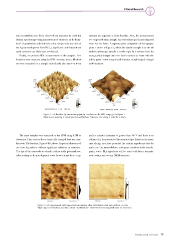
non-repeatability have been observed and discussed in detail for vacuum and represent a clean baseline. Next, the measurements
Raman spectroscopy using nanostructured substrates in the litera- were repeated with a sample that was submerged in non-degassed
14
ture . Hypothesized to be intrinsic to the non-periodic structure of water for two hours. A representative comparison of the topogra-
the Ag nanorods grown from PVD, a significant contribution from phies is shown in Figure 5, where the baseline sample is on the left
rapid corrosion has likely been overlooked. and the submerged sample is on the right. It is evident from the
Finally, we present SPM measurements of the samples. Five topographical images that even brief exposure to water with dis-
locations were measured using the SPM in contact mode. The first solved gases results in rapid and dramatic morphological changes
set were measured on a sample immediately after removal from to the surfaces.
Figure 5: Left: Baseline Ag nanorods topography correlative to the SEM imaging A in Figure 1.
Right: Local mapping of topography for Ag Oxidized Nanorods after sitting in water for 2 hours.
The same samples were analyzed on the SPM using KFM to surface potential increases to greater than 3.0 V and there is no
determine if the surfaces have chemically changed from the base- evidence for the presence of fine nanorod tips. Based on the meas-
line state. The baseline, Figure 6 left, shows the potential measured ured change in surface potential, the authors hypothesize that the
on clean Ag surfaces without significant oxidation or corrosion. surfaces of the nanorods have undergone oxidation in the non-de-
The tips of the nanorods are clearly evident in the potential plot. gassed water. This hypothesis will be tested with future transmis-
After soaking in the non-degassed water for two hours the average sion electron microscopy (TEM) analyses.
Figure 6: Left: Ag nanorods surface potential contour map taken immediately after removal from vacuum.
Right: Ag nanorod surface potential contour map taken after submersion in non-degassed water for two hours.
Shimadzu Journal vol.9 Issue1 17