
Page 9 - Shimadzu Xslicer SMX-6010
P. 9
Versatile, User-Friendly Functions
Teaching Function Panoramic Imaging Function
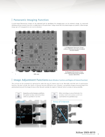
Fluoroscopic and CT imaging can be automated using the Teaching Function, which moves the sample stage to preregistered A wide-range fluoroscopic image can be obtained just by specifying the imaging span on the external image. An improved
points of interest. stitching process ensures there are no visible lines in the panoramic image where the individual images are joined. A fluoroscopic
Additionally, for visual inspection, OK and NG judgment functions are included. image up to 32 megapixels in size can be obtained.
Point 1 (Fluoroscopic imaging)
2.23 Megapixel Panoramic Image
Point 2 (CT imaging) without Stitching (Partially Enlarged)
300 mm
(Former Model)
Point 3 (Fluoroscopic imaging)
Point 5 (Fluoroscopic imaging) Point 4 (CT imaging)
32 Megapixel Panoramic Image
with Stitching (Partially Enlarged)
300 mm (Xslicer SMX-6010)
Stepwise Movement
The Stepwise Movement function moves the stage at constant intervals. It specifies the starting position, amount of movement, Image Adjustment Functions (Auto Window Function and Region of Interest Function)
and number of movements. When this function is used, observations are performed while the stage makes consecutive movements
from the starting position in accordance with the settings. Consecutive fluoroscopic or CT scans can be performed of samples The contrast can be automatically optimized to make the area of interest easy to see. Normally, with this sort of optimization
arranged at set intervals.
function, the part outside the region of interest becomes difficult to see. However, a proprietary image processing algorithm
automatically adjusts the image to ensure that the part outside the region of interest remains as easy to see as possible.
Example of Consecutive CT Images of
25 Samples Aligned on a Pallet
Depending on the changing conditions, When selecting an area of interest, the
the optimal window level and window window level and window width are
width are specified in real time. optimized for the specified range.
Cross-Sectional Image
AW ON Area of Interest
ICs Aligned on a Pallet
Continuous CT scan possible Determination Exterior
(e.g., 25 scans) Results Image
AW OFF
A list of the determination results is displayed when
all the configured points have been inspected.
Selective measures can be performed after inspecting the results.
Xslicer SMX-6010
8 Microfocus X-Ray Inspection System 9