
Page 9 - LifeScience Solution for FNIRS
P. 9
For Research Use Only. Not for use in diagnostic procedures.
Neurorehabilitation Comparison of fNIRS and fMRI Light to Measure Brain Function Principle of Using Near Infrared
Example of Brain-Function Imaging Research: Neurorehabilitation Application Research Example of Brain-Function Imaging Research: Comparison of fNIRS and fMRI
Data Data Imaging Optical Brain-Function
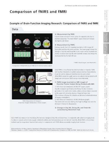
In recent years, progress has been made in researching the use of fNIRS in neurorehabilitation applications, such as during or after a A: Measurement by fMRI
stroke. A B
PET and fMRI systems require the subject to be at rest, but fNIRS systems can measure brain function even while the subject is Clearly shows activity in motor cortex for opposite side due to bilitation Neuroreha-
exercise (grasping). The lower BOLD* signal shows an increase
performing tasks that involve body movement. Therefore, it can be used to obtain information about cerebral activity associated with
during the task (blue).
walking or other movement.
B: Measurement by fNIRS
Irradiation
Displays results from 2D mapping (overlaid on MRI image) of
Detection Deoxy-Hb and Oxy-Hb during exercise. The lower graph shows the fNIRS and fMRI Comparison of
change in Oxy-Hb and Deoxy-Hb in the motor cortex (marked with
circles). It shows how Oxy-Hb and Total-Hb increase and Deoxy-Hb
decreases during the task (40 sec).
Walk Walk Walk Walk Walk Motor Control Brain Activity during
* BOLD: Blood Oxygen Level Dependent
Comparison of BOLD-fMRI (A) and fNIRS (B) for Normal Healthy Subject
Since fNIRS is not affected by electrical noise or magnetic fields
C D Measurement with EEG Simultaneous
and has few limitations on patient posture during measurements,
fNIRS Measurement of Brain Activation while Walking on a Treadmill
it can be used to measure brain function in cases where
BOLD-fMRI cannot be used, such as on subjects being treated with
Walk Swinging Arms Leg Movement Visualizing Walking Tremor(+) deep brain stimulation (DBS) using a metal electrode.
C: fNIRS Image (overlaid on MRI image) of Analysis Method NIRS Signal
Essential Tremor Subject Being Treated with DBS
1. With Electrical Stimulation of DBS OFF (upper graph)
Tremor(-) Oxy-Hb increased significantly and Deoxy-Hb also increased,
fNIRS
primarily in the right motor cortex, when a strong tremor occurred
in the upper left extremity during a finger-to-finger test. Inner Speech Investigating
(The increase in Deoxy-Hb indicates an abnormal enhanced release
of oxygen metabolites.)
2. With Electrical Stimulation of DBS ON (lower graph)
fNIRS Measurement of Change in The tremor did not occur during the finger-to-finger test and the
Active Brain Oxygen Metabolites in Tremor Subject
increase in Deoxy-Hb disappeared. Functions Language Processing
fMRI
D: Change in Oxy-Hb, Deoxy-Hb, and Total-Hb
Levels During Movement Task
Brain Activity for Healthy Person during walking and Related Tasks
The above shows the brain activity measured while walking on a treadmill. It shows an increase in Oxy-Hb associated with walking, Research Mental Disorder
Since fNIRS can measure not only Deoxy-Hb, but also changes in Oxy-Hb and blood flow, it is especially well suited to imaging brain
inner side of the primary sensorimotor cortex, near the center. fNIRS is able to measure brain activity during dynamic movements
function in subjects where brain oxygen metabolite activity and hemodynamics are not normal. Currently, BOLD-fMRI is the primary
assigned as tasks, such as walking or arm swinging, which cannot be measured using fMRI and PET. It can also be used to assess
method used to image brain function, but using it in conjunction with fNIRS allows accurately imaging brain function.
brain activity at the bedside. fNIRS is being used in research to evaluate brain activity during hemiplegic gait by stroke subjects, to
improve asymmetry in the activity of the sensorimotor cortex, increase activity in the premotor cortex, improve walking, etc. Shimadzu fNIRS Key References Regarding
(Data provided by: Mr. Ichiro Miyai, Morinomiya Hospital, Omichi-kai Medical Corporation) (Data provided by: Mr. Kaoru Sakatani, Department of Neurological Surgery, Nihon University School of Medicine)
Reference: Miyai, I., (2004). "Application of fNIRS in Neurorehabilitation." MEDICAL NOW, No. 52: 33-36. Reference: Sakatani, K., (2006) "Imaging Brain-Function of Patients with Encephalopathy: Comparison of fNIRS and fMRI," MEDICAL NOW, No.59: 44-46.
8 9