Page 34 - Pharmaceutical- Guide to Biopharmaceutical
P. 34

Characterization                Quality Control


                     Quantitation of Proteins                                                                                           BioSpec-nano


                                       Quantitation of Proteins Using                                                                                      benefits

                                                      BioSpec-nano                                                                                                                                                                           Cell Line Optimization
                                                                                                                                                •  Measure the concentration of proteins or check the purity.
                                                                                                                                                •  Measure sample quantities as small as 1 μL.
                     Operating Principle and Features
                                                                                                                                                •  Achieve low carryover with the automatic wiping function.
                     The BioSpec-nano has two available optical path lengths, 0.2 mm and
                     0.7 mm, and can quantify proteins and nucleic acids in sample quantities
                     as low as 1 µL. Samples can also be measured using an optional cell with                                                                                                                                                Culture
                     a 5 mm optical path length (for 2 mL sample quantities). That means
                     only a small sample quantity is needed for quick protein concentration
                     measurements or to check spectra.
                     A wiping mechanism enables automatic cleaning between samples,
                     ensuring extremely low carryover and reducing inconsistencies that
                     may occur with manual cleaning (Fig. 1).
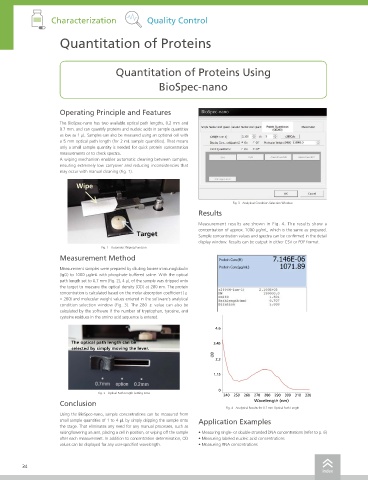
                          Wipe


                                                                              Fig. 3   Analytical Condition Selection Window                                                                                                                 Purification
                                                                   Results

                                                                   Measurement results are shown in Fig. 4. The results show a
                                                                   concentration of approx. 1000 μg/mL, which is the same as prepared.
                                              Target               Sample concentration values and spectra can be confirmed in the detail
                                                                   display window. Results can be output in either CSV or PDF format.
                                   Fig. 1   Automatic Wiping Function
                     Measurement Method                                                                                                                                                                                                      Characterization

                     Measurement samples were prepared by diluting bovine immunoglobulin
                     (IgG) to 1000 μg/mL with phosphate buffered saline. With the optical                                               Specifications
                     path length set to 0.7 mm (Fig. 2), 4 μL of the sample was dripped onto
                     the target to measure the optical density (OD) at 280 nm. The protein                                               Instrument            BioSpec-nano
                     concentration is calculated based on the molar absorption coefficient (ε
                     = 280) and molecular weight values entered in the software's analytical                                             Wavelength range      220 to 800 nm
                     condition selection window (Fig. 3). The 280 ε value can also be                                                    Spectrum bandwidth    3 nm
                     calculated by the software if the number of tryptophan, tyrosine, and
                     cysteine residues in the amino acid sequence is entered.                                                            Wavelength accuracy   ±1 nm                                                                         Quality Control
                                                                                                                                         Pathlength            0.2 mm, 0.7 mm
                                                                        4.6
                                                                                                                                         Photometric value unit  OD (Optical Density), absorbance converted with 10 mm pathlength
                         The optical path length can be                 3.45                                                             Sample volume         1 µL min. (pathlength: 0.2 mm)
                                                                                                                                                               2 µL min. (pathlength: 0.7 mm)
                         selected by simply moving the lever.
                                                                                                                                         Light source          Xenon flash lamp
                                                                        2.3
                                                                                                                                         Monochromator         Holographic grating
                                                                                                                                         Detector              Photo diode array
                                                                        1.15
                                                                                                                                         Auto wiping function  Provided                                                                      Pharmacokinetics
                                                                                                                                         Spectrum measuring time  3 sec
                                                                         0
                                  Fig. 2   Optical Path Length Setting Area
                                                                           240  250  260  270  280  290  300  310  320                   Quantitation range    Pathlength 0.2 mm, 1 to 75 OD, 50 to 3,700 ng/µL
                     Conclusion                                                      Wavelength (nm)                                                           Pathlength 0.7 mm, 0.3 to 21 OD, 15 to 1,000 ng/µL
                                                                            Fig. 4   Analytical Results for 0.7 mm Optical Path Length                         Optional 5 mm pathlength cell, 0.04 to 3 OD, 2 to 150 ng/μL
                     Using the BioSpec-nano, sample concentrations can be measured from                                                  Dimensions            W 210 mm x D 214 mm x H 417 mm
                     small sample quantities of 1 to 4 μL by simply dripping the sample onto   Application Examples
                     the stage. That eliminates any need for any manual processes, such as                                               Weight                7 kg                                                                          Others
                     raising/lowering an arm, placing a cell in position, or wiping off the sample   • Measuring single- or double-stranded DNA concentrations (refer to p. 6)  Analysis mode  Simple nucleic acid quantitation, labeled nucleic acid quantitation, protein quantitation,
                     after each measurement. In addition to concentration determination, OD   • Measuring labeled nucleic acid concentrations                  labeled protein quantitation, photometric measurement
                     values can be displayed for any user-specified wavelength.  • Measuring RNA concentrations                         Note: The droplet formation status will affect analysis results. Measure quantities that are large enough to enable proper droplet formation.


        34                                                                                                                                                                                                                                  35
                                                                                                            index                                                                                                                  index
   29   30   31   32   33   34   35   36   37   38   39