
Page 12 - Pharmaceutical- Guide to Biopharmaceutical
P. 12
Culture
Metal Elements Easily Quantified during Culturing AA-7000 Series
Monitoring of Metal Elements in Cell Culture benefits
Supernatant using Atomic Absorption Spectrometry Cell Line Optimization
click here • Metal elements in culture media can be analyzed without any complicated pretreatment steps.
• Multiple trace metal elements can be quantified inexpensively and easily.
Operating Principle and Features Table 1 Comparison of Atomization Methods
Electric thermal method Flame method • The system supports the electric thermal method, flame method, or automatically
Atomic absorption spectrometry involves atomizing elements at high Sensitivity ppt to ppb ppb to ppm switching between the methods.
temperature to quantitate element concentrations based on the absorption Atomization efficiency 90 % or more Approx. 10 %
of specific light wavelengths during atomization. Required sample/analysis 5 to 50 µL 1 to 2 mL Culture
2 to 5 min
5 to 10 sec
There are two main atomization methods: (1) the electric thermal method, Analysis time/analysis RSD 3 % (approx.) RSD 1 % (approx.)
Repeatability
which involves generating heat with an electrical current (high sensitivity),
or (2) the flame method, which involves heating with a flammable gas Table 2 Analytical conditions of the electric thermal method
flame. (Table 1 shows a comparison.) Either method can be used in AA- Analysis Slit width Atomization Lighting
7000 systems, which include an auto-atomizer changer (AAC) that can wavelength (nm) Ashing temp. temp. mode Tube type
(nm)
be used to automatically switch between the methods for measurements.
Cu 324.8 0.7 2500 °C
Mn 279.5 2200 °C
Measurement Method and Conditions Co 240.7 0.2 800 °C 2300 °C BGC-D2 Platform tube
Fe 248.3 2300 °C
The high concentrations of Mg and Zn were measured using the flame Purification
method and trace elements (Cu, Mn, Co, and Fe) using the electric thermal Table 3 Analysis conditions of the flame method
method, based on the analytical conditions indicated in Tables 2 and 3. Analysis
CHO cells were inoculated in a 125 mL flask and cultivated by shaking for wavelength Slit width (nm) Lighting mode Flame type C2H2 flowrate
(nm)
four days. Every 24 hours, from immediately after starting cultivation, 1 Zn 213.9 2.0 L/min
mL of the cell culture fluid was sampled, removed cells by centrifugation, Mg 285.2 0.7 BGC-D2 Air ー Acetylene 1.8 L/min Specifications
and then the supernatant was collected. Samples were diluted by 20
times for Cu, Mn, and Zn, 40 times for Co and Fe, and 500 times for Mg Abs Abs Instrument AA-7000F/AAC
before analysis (nitric acid was diluted to 0.5 v/v%). Standard solutions for Cu_Day 3 Fe_Day 3
each element were prepared by diluting the standard solution for atomic - : Standard of 3 ppb - : Standard of 20 ppb Wavelength range 185.0 to 900.0 nm
- : Spiked sample
- : Spiked sample
absorption spectrometry (1000 mg/L). The nitric acid concentration was - : Culture supernatant - : Culture supernatant Bandwidth 0.2, 0.7, 1.3, 2.0 L nm (4-step automatic switching)
- : BG
- : BG
prepared to 0.5 v/v%. The calibration curve method was used for all Characterization
analyses. Background correction BGC-SR (high-speed self-reversal method) (185.0 to 900.0 nm), BGC-D2 (D2 lamp method) (185.0 to
method 430.0 nm)
Results Lamp mode EMISSION, NON-BGC, BGC-D2, BGC-SR
Measurement mode Flame continuous method, flame micro sampling method, furnace method, flame emission method
The calibration curve coefficient of correlation was r = 0.999 or higher for
all components. A spike-and-recovery test was performed for each element Maximum reagent / Reagents: 8 positions, Samples: 60 positions (when using an autosampler)
by adding a standard solution with a fixed concentration. (The additive sample positions
recovery rate equals the concentration difference between spiked and Digital recording Management by login ID and password, control user access authority by user level, log record, audit
unspiked samples divided by the additive concentration.) Test results were trail, electronic signatures Quality Control
roughly within 100 ±10 %, which is an excellent additive recovery rate. Positioning Automatic flame/furnace switching by motor
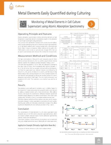
The electric thermal method and flame method were also used to monitor Sec Sec
time-series changes in culture supernatant concentrations for each sample. Fig. 1 Peak profile Dimensions and weight W 700 × D 588 × H 714 mm, 76 kg (Autosampler is not included.)
Resulting peak profiles and time-series concentration changes in the culture Burner head Titanium 10 cm slot (5 cm titanium slot for N2O–C2H2 flame available as an option)
supernatant obtained by the two methods are shown in Figs. 1 and 2. 20 Mn Nebulizer Pt-lr capillary, PTFE orifice, ceramic impact bead (capable of handling hydrofluoric acid)
Conclusion 10 11.9 8.84 16.9 Flame Type Air–C2H2, N2O–C2H2
Conc. (µg/L) 11.1 11.5 Safety measures Automatic gas leak check, automatic Air–N2O switching as C2H2 flowrate increases, flame monitor,
The concentrations of metal elements in a cell culture supernatant were prevention of wrong burner head use, gas pressure monitor, drain tank level monitor, automatic flame
measured using an AA-7000 atomic absorption spectrophotometer, which 0 extinction upon power outage or sudden power interruption, automatic flame extinction via flame Pharmacokinetics
can measure samples using two types of atomic absorption spectrometry Day 0 Day 1 Day 2 Day 3 Day 4 vibration sensor, internal fan stop sensor
methods, electric thermal and flame. 60 Heating control system Drying: Digital current control with automatic temperature calibration function
Time-course changes in metal element concentrations can be monitored Mg Ashing, Atomization: Digital temperature control via optical sensor
using only a simple pretreatment step of diluting the cell culture supernatant. 50 53.3 50.1 48.0 47.9 47.4 Carryover Rinse port: Less than 0.0001 Mixing port: Less than 0.00001
Application Example (Shimadzu Application News No.) Conc. (mg/L) Furnace Auto dilution / re-analysis For measurement result on unknown samples
· If extrapolation of calibration curve is possible: automatic calculation of dilution rate and dilution to
• Analysis of metallic elements in cell culture medium (A634) 40 bring concentration within calibration curve range Others
Day 0 Day 1 Day 2 Day 3 Day 4 · If extrapolation of calibration curve is not possible: dilution rate fixed at 10×
Note: Value obtained by converting the measurement value to one corresponding to stock solution of cell culture supernatant. Safety measures Cooling water flowrate monitor, gas pressure monitor, overcurrent protection unit (double check by
circuit protector and optical sensor), furnace block cooling check
Fig. 2 Time course of Mn and Mg concentration in culture supernatant
12 13
index index