
Page 9 - Shimadzu Journal vol.2 Issue2
P. 9
New Technology 67
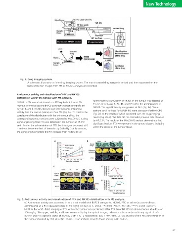
significant levels of PTX were present in the tumour clusters, including
results (Fig. 2b–e). The data did not contradict previous data obtained
following the accumulation of NK105 in the tumour was detected at
sections serial to those for MALDI-MS were also quantified by LCMS
by HPLC14. The results of the MALDI-MS analysis demonstrate that
15 min as well as at 1, 24, 48, and 72 h after the administration of
(Fig. 2d, e), the results of which correlated with the drug imaging
NK105. The signal intensity was greatest at 24 h (Fig. 2c). Tissue
A schematic illustration of the drug imaging system. The matrix-coated drug sample is ionised and then separated on the
ɹɹɹ
within the centre of the tumour tissue. (a) Antitumour activity was examined in an animal model with BXPC3 xenografts. NK105, PTX, or saline (as a control) was administered at a PTX equivalent dose of 50 mg/kg on days 0, 4, and 8. *P< 0.05 (PTX vs. NK105), ***P< 0.001 (saline vs. NK105). Bar = SD. (b)(c) Imaging of PTX within the tumour was performed after PTX (b) or NK105 (c) administration at a dose of 100 mg/kg. The upper, middle, and lower columns display the optical images, reference substance (an arbitrary signal of m/z 824.6), and PTX (specific signal of m/z 892.3 [M + K] + ), respectively. Bar, 1 mm. (d)(e) LC-MS a
system. basis of its m/z. Images from MS or MS/MS analysis are recorded. Antitumour activity and visualisation of PTX and NK105 distribution within the tumour with MS analysis. NK105 or PTX was administered at a PTX equivalent dose of 50 mg/kg/day to mice bearing BxPC3 pancreatic cancer xenografts on days 0, 4, and 8. NK105 showed significantly higher antitumour activity than the control (saline) and free PTX (Fig. 2a). To confirm the correlation of the distribution with the antitumour effect, the corresponding tumour sections were subjected to MALDI-IMS. A drug signal originating from PTX was detected in the tumours at 15 min and 1 h after the administration of
imaging
Drug
1.
Fig.
with that of PTX alone after injection into tumour-bearing mice. We demonstrated optically and quantitatively that NK105 delivered more PTX to the
spectrometry (MALDI-IMS) with enhanced resolution and sensitivity, we compared the distribution of a paclitaxel (PTX)-incorporating micelle (NK105)
treatment yielded a greater antitumour effect and less neural toxicity in mice than did PTX treatment. The use of high-resolution MALDI-IMS may be
Masahiro Yasunaga 1 , Masaru Furuta 2 , Koretsugu Ogata 2 , Yoshikatsu Koga 1 , Yoshiyuki Yamamoto 1 , Misato Takigahira 1 & Yasuhiro
In this study, we investigated the ability of our mass microscopy technique
desorption/ionisation and quadruple ion trap time-of-flight (TOF) analyser.
We have developed a mass microscopy method in which a microscope is
setting are desirable for evaluating drug effects and optimising drug design. Here, using matrix-assisted laser desorption ionisation imaging mass
1 Division of Therapeutics Development, Research Center for Innovative Oncology, National Cancer Center Hospital East, 6-5-1
tumour, including the centre of the tumour, while delivering less PTX to normal neural tissue, compared with injection with PTX alone. NK105
formulation and obtain precise regional information about the drug
to visualise the tissue distribution of unlabelled ACA and its micellar
2 Analytical & Measuring Instruments Division, Shimadzu Corporation, 1, Nishinokyo-Kuwabaracho, Nakagyo-ku, Kyoto
coupled with a high-resolution atmospheric pressure-laser
distribution in a specific anatomical area. Results The drug imaging system and its application in PTX analysis on the MALDI target. A schematic representation of our drug imaging system is shown in Fig. 1. Imaging data were acquired using a mass microscope. In the analysis, mass spectrometry (MS) and tandem mass spectrometry (MS/MS) were used for quantification and validation, respectively (Fig. 1). Paclitaxel (PTX) is a mitotic inhibitor and an ACA that is used to treat various cancers. However, PTX is associated with peripheral neuropathy, a serious adverse affect 13 . NK105, a PTX-incorporating micelle, was developed to
global w430×h280 The significance of microscopic mass spectrometry with high resolution in the visualisation of drug distribution Kashiwanoha, Kashiwa, Chiba 277-8577, Japan, Correspondence and requests for materials should be addressed to Y.M. (yhmatsum@east.ncc.go.jp) The visualisation and quantitative analysis of the native drug distribution in a pre-clinical or clinical an innovative approach for pharmacological evaluation and drug design support. Drug delivery, Pharmacokinetics, Pharmacodynamics, Biochemical assays Advances in our understanding of cancer at the cellular and molecular levels have promoted the development of new drugs 1,2 . Pharmacokinetic (PK) and pharmacodynamic (PD) studies are very important t
New Technology Matsumura 1 604-8511, Japan. ABSTRACT Key Words Introduction urgently required. 66