Page 8 - Shimadzu SPM-8100FM
P. 8

Examples of Hydration/Solvation Structure Measurements                                                                    The Principle of FM-Type AFM


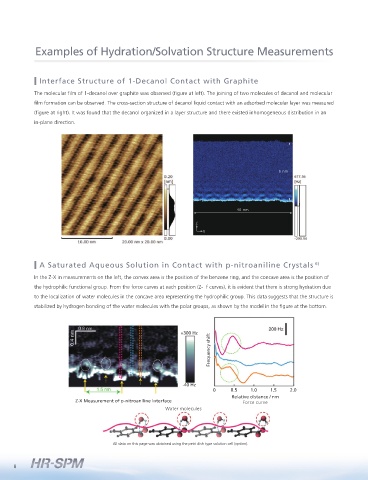
                     Interface Structure of 1-Decanol Contact with Graphite                                                                   The frequency of a vibrating cantilever is measured in dynamic mode, and interactions with the sample are detected.
                                                                                                                                              Specifically, the cantilever is moved in a non-contact state, so that the cantilever frequency shift (  f ) is constant.
                    The molecular film of 1-decanol over graphite was observed (figure at left). The joining of two molecules of decanol and molecular
                                                                                                                                              This enables highly sensitive force detection, 20 times better than with existing methods, thereby improving the
                    film formation can be observed. The cross-section structure of decanol liquid contact with an adsorbed molecular layer was measured
                                                                                                                                              image resolution.
                    (figure at right). It was found that the decanol organized in a layer structure and there existed inhomogeneous distribution in an
                    in-plane direction.
                                                                                                                                                                                                                 Cantilever resonance curve

                                                                                                                                                                                                                          f
                                                                                                                                                        Probe           Cantilever
                                                                                                                                                                    (Dynamic resonance system)
                                                                                                                                                                  Sample
                                                                                                                                                                                            Cantilever      Oscillation amplitude
                                                                                                     6 nm
                                                                                                                                                                           Interaction force
                                                                                                                                                                            (attraction)
                                                                                                                                                                            Non contact


                                                                                                                                                                                                                      Frequency
                                                                                       40 nm
                                                                         Z
                                                                                                                                                References
                                                                            X
                                                                                                                                                Cited References
                                                                                                                                                1) Ryohei Kokawa, Masahiro Ohta, Akira Sasahara, Hiroshi Onishi, Kelvin Probe Force Microscopy Study of a Pt/TiO 2 Catalyst Model Placed in an Atmospheric Pressure of N 2
                                                                                                                                                  Environment, Chemistry – An Asian Journal, 7, 1251-1255 (2012).
                     A Saturated Aqueous Solution in Contact with p-nitroaniline Crystals                        6)                             2) K. Nagashima, M. Abe, S. Morita, N. Oyabu, K. Kobayashi, H. Yamada, R. Murai, H. Adachi, K. Takano, H. Matsumura, S. Murakami, T. Inoue, Y. Mori, M. Ohta, R. Kokawa, Molecular
                                                                                                                                                  resolution investigation of tetragonal lysozyme(110) face in liquid by FM-AFM, Journal of Vacuum Science and Technology B 28 (2010) C4C11-C4C14
                    In the Z-X in measurements on the left, the convex area is the position of the benzene ring, and the concave area is the position of   3) Sebastian Rode, Noriaki Oyabu, Kei Kobayashi, Hirofumi Yamada, and Angelika Kuhnle, True Atomic-Resolution Imaging of  (1014) Calcite in Aqueous Solution by Frequency
                    the hydrophilic functional group. From the force curves at each position (Z-  f curves), it is evident that there is strong hydration due   Modulation Atomic Force Microscopy, Langmuir,  2009,  25 (5),  pp 2850–2853
                                                                                                                                                4) K. Kimura, S. Ido, N. Oyabu, K. Kobayashi, Y. Hirata, T. Imai, H. Yamada, Visualizing water molecule distribution by atomic force microscopy, Journal of Chemical Physics, 132, 19,
                    to the localization of water molecules in the concave area representing the hydrophilic group. This data suggests that the structure is
                                                                                                                                                  194705 (2010).
                    stabilized by hydrogen bonding of the water molecules with the polar groups, as shown by the model in the figure at the bottom.  5) Kei Kobayashi, Noriaki Oyabu, Kenjiro Kimura, Shinichiro Ido, Kazuhiro Suzuki, Takashi Imai, Katsunori Tagami, Masaru Tsukada and Hirofumi Yamada, Visualization of hydration
                                                                                                                                                  layers on muscovite mica in aqueous solution by frequency-modulation atomic force microscopy, Journal of Chemical Physics, 138, 184704 (2013).
                                                                                                                                                6) Rina Nishioka, Takumi Hiasa, Kenjiro Kimura, and Hiroshi Onishi, Specific Hydration on p Nitroaniline Crystal Studied by Atomic Force Microscopy, J. Phys. Chem. C, 117, 2939−
                                                                                                                                                  2943 (2013).
                                                                    +300 Hz                                                                     Instrument References
                                                                              Frequency shift                                                    • Kei Kobayashi, Hirofumi Yamada, and Kazumi Matsushige, Reduction of frequency noise and frequency shift by phase shifting elements in frequency modulation

                                                                                                                                                 • Ryohei Kokawa and Masahiro Ohta, Development of a High Resolution FM-AFM Working in Air or Solution, Microscopy, Vol. 47, No. 1 (2012) (Japanese)

                                                                                                                                                  atomic force microscopy, Rev. Sci. Instrum., 82, 033702 (2011).

                                                                                                                                                Application Examples
                                                                     -40 Hz
                                                                               0     0.5   1.0    1.5   2.0                                      • Shinichiro Ido, Kenjiro Kimura, Noriaki Oyabu, Kei Kobayashi, Masaru Tsukada, Kazumi Matsushige and Hirofumi Yamada, Beyond the Helix Pitch: Direct
                                                                                     Relative distance / nm                                       Visualization of Native DNA in Aqueous Solution, ACS Nano, 7, 1817-1822 (2013).
                                 Z-X Measurement of p-nitroaniline interface             Force curve                                             •  Takumi  Hiasa  and  Hiroshi  Onishi,  Competitive  Adsorption  on  Graphite  Investigated  Using  Frequency-Modulation  Atomic  Force  Microscopy:  Interfacial  Liquid
                                                               Water molecules                                                                    Structure Controlled by the Competition of Adsorbed Species, Langmuir, 29, 5801-5805 (2013).


                                                                                                                                                This Instrument has been commercially developed through collaboration with the Yamada group at Kyoto University, the Morita group at Osaka University, the Onishi group at
                                                                                                                                                Kobe University, the Tomitori group at JAIST, and the Arai group at Kanazawa University.
                                              All data on this page was obtained using the petri dish type solution cell (option).

                                                                                                                                                                                                                                SPM-8100FM
             8                                                                                                                                                                                                                High Resolution Scanning Probe Microscope  9
   3   4   5   6   7   8   9   10   11   12