
Page 6 - Shimadzu iMScope QT
P. 6
Combined Analysis
Fusion of observations from an optical microscope with MS images
(exclusive to Shimadzu)
MS images can be obtained exibly and matched to observation images,
either the entire image area or detailed portions of it.
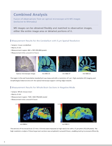
Measurement Results for the Cerebellum with 5 m Spatial Resolution
• Sample: mouse cerebellum
• Matrix: 9-AA
• Measurement region: 662 × 595 (393,890 pixels)
• Measurement time: around 2.2 hours
Optical microscope image m/z 885.55 m/z 888.57 m/z 888.63
The region in the red frame below (cerebellum) was measured with a resolution of 5 m. High-resolution MS imaging and
morphological observations with the optical microscope support cutting-edge research.
Measurement Results for Whole Brain Sections in Negative Mode
• Sample: Whole mouse brain
• Matrix: 9-AA
• Measurement region: 1126 × 624 (702,624 pixels)
• Measurement time: around 6 hours
m/z 888.6 m/z 885.5
The sections of the mouse brain (17 mm × 9.4 mm) were measured at high resolution with a 15 m pitch (702,624 pixels). The
high-resolution analysis of these large brain sections was completed in around 6 hours, enabling testing to proceed ef ciently.
6