
Page 17 - Shimadzu EPMA-1720 Series
P. 17
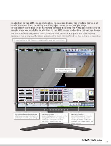
In addition to the SEM image and optical microscope image, the window controls all
             hardware operations, including the X-ray spectrometer and sample stage.
             In the observation display, all hardware controls including the X-ray spectrometer and
             sample stage are available in addition to the SEM image and optical microscope image.
             The user interface is designed to reveal the status of all hardware at a glance and offer intuitive
             operation. Frequently used functions appear on the front window for stress-free instrument operation.

                Common functions for SEM observations are arranged on upper and lower toolbars.


                                                              Simultaneous display of SEM and
                                                              optical microscope images


                  Electron optical system control window  WDS control window  Stage control window
                  Offers detailed display and adjustment   Graphic display shows X-ray spectrometer   Simultaneous display of the overall
                  of the electron optical system settings.   settings for each channel at a glance.   map and enlarged map created
                                               Controls are arranged for intuitive   according to the mounted holder
                                               operation of required functions.   allows accurate identification of the
                                                                              current position.


                                                                                           EPMA-1720 Series
                                                                                                   Electron Probe Microanalyzer  17