Page 8 - Shimadzu Xslicer SMX-1010
P. 8

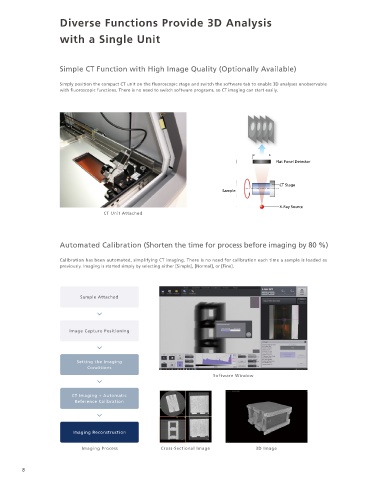
Diverse Functions Provide 3D Analysis


                     with a Single Unit


                     Simple CT Function with High Image Quality (Optionally Available)                                                  Panoramic Imaging Function


                     Simply position the compact CT unit on the fluoroscopic stage and switch the software tab to enable 3D analyses unobservable   A wide X-ray fluoroscopic image can be obtained just by specifying the imaging range on the exterior image. An improved
                     with fluoroscopic functions. There is no need to switch software programs, so CT imaging can start easily.          stitching process ensures a panoramic X-ray fluoroscopic image up to 32 megapixels in size can be obtained with no conspicuous
                                                                                                                                        marks where the images are spliced together.

                                                                                                                                                                    300 mm


                                                                                            Flat Panel Detector                                                                                        Panoramic Image
                                                                                                                                                 300 mm                                           No Splicing (region enlarged)


                                                                                              CT Stage
                                                                           Sample

                                                                                              X-Ray Source
                                   CT Unit Attached


                                                                                                                                                            32 Megapixel Panoramic Image               Panoramic Image
                     Automated Calibration (Shorten the time for process before imaging by 80 %)                                                      Combined 9 × 10 images by Xslicer SMX-1020   Splicing (region enlarged)

                                                                                                                                                                                                             300 x 350 mm
                     Calibration has been automated, simplifying CT imaging. There is no need for calibration each time a sample is loaded as   Imaging Mode              Pixels                       Scanning time for entire range
                     previously. Imaging is started simply by selecting either [Simple], [Normal], or [Fine].
                                                                                                                                            Simple    Equivalent to 2K (full HD) in size  Approx. 2 million pixels  SMX-1010 : 115 sec / SMX-1020 : 100 sec


                                                                                                                                            Normal        Equivalent to 4K in size  Approx. 8 million pixels  SMX-1010 : 135 sec* / SMX-1020 : 120 sec
                            Sample Attached
                                                                                                                                             Fine         Equivalent to 8K in size  Approx. 32 million pixels  SMX-1010 : 590 sec / SMX-1020 : 495 sec

                                                                                                                                                                                                  *The SMX-1000 Plus takes 395 seconds at the fastest.
                        Image Capture Positioning
                                                                                                                                        Image Adjustment Functions
                                                                                                                                        (Auto Window Function and Area of Interest Function)

                           Setting the Imaging                                                                                          The contrast can be automatically optimized to make the area of interest easy to see.
                              Conditions
                                                                        Software Window
                                                                                                                                                         AW ON                                          Area of Interest

                         CT Imaging + Automatic
                          Reference Calibration
                                                                                                                                        The optimal window                             When selecting an
                                                                                                                                        level and window                               area of interest, the
                                                                                                                                        width are configured   AW OFF                   window level and
                                                                                                                                        in real time even if the                       window width are
                         Imaging Reconstruction                                                                                         conditions change.                             optimized for the
                                                                                                                                                                                       specified range.
                            Imaging Process           Cross-Sectional Image           3D Image

                                                                                                                                                                                                            Xslicer SMX-1010/1020
        8                                                                                                                                                                                                                 Microfocus X-Ray Inspection System  9
   3   4   5   6   7   8   9   10   11   12   13