
Page 7 - Shimadzu Xctal 5000
P. 7
Dual Observation Method for
Absorption and Phase Applications (Phase Images)
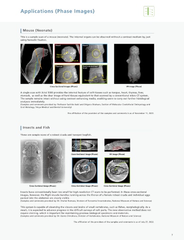
High-Contrast Observations Mouse (Neonate)
by Absorption Images and Phase Images This is a sample scan of a mouse (neonate). The internal organs can be observed without a contrast medium by, just
using formalin fixation.
A phase image is suited to observations of samples with large density differences. Head Heart
In contrast, an absorption image is suited to observations of samples with large differences in absorption coefficient.
The X-ray absorption coefficient makes it possible to visualize samples with no contrast differences, as long as there
are density differences. Using these two observation methods widens the scope of observations by enabling Nasal
high-contrast sample observations. septum
Tongue
Thymus Gastrointestinal
Water and Acrylic tract
The density difference is larger than the absorption coefficient difference, enabling high-contrast phase image
observations. Stomach Liver
High-contrast phase image observations are possible if the density difference is approximately 0.2 g/cm³ or larger.
Cross-Sectional Images (Phase) VR Image (Phase)
Acrylic Water
A single scan with Xctal 5000 provides the internal feature of soft tissues such as tongue, heart, thymus, liver,
stomach, as well as the clear image of hard tissues equivalent to that scanned by a conventional micro CT system.
Absorption
Coefficient 0.36 0.38 The sample remains intact without using contrast enhancing media, enabling users to carry out further histological
cm - ¹ analyses immediately.
(Samples and comments provided by: Professor Sachiko Iseki and Shigeru Okuhara, Section of Molecular Craniofacial Embryology and
Density 1.19 1 Oral Histology, Tokyo Medical and Dental University)
g/cm³
The affiliation of the providers of the samples and comments is as of November 11, 2022.
Cross-Sectional Image Cross-Sectional Image
(Absorption) (Phase) Insects and Fish
These are sample scans of a robust cicada and twospot hogfish.
Water and Multiple Types of Resins
The contrast differs for the absorption image and the phase image. As shown in the graphs below, it is evident that
the brightness value for the absorption image is proportional to the absorption coefficient, and the brightness value
for the phase image is proportional to the density.
Cross-Sectional Image (Phase) VR Image (Phase)
PVC PVC ABS PVC PTFE PE POM Water
ABS ABS Brain Internal organs
PTFE PTFE Absorption
Coefficient 0.29 1.87 0.87 0.26 0.47 0.38
cm - ¹
POM POM Density
PE PE g/cm³ 1.07 1.26 2.17 0.96 1.42 1
Cross-Sectional Image Cross-Sectional Image Cross-Sectional Image (Phase) Cross-Sectional Image (Phase) Cross-Sectional Image (Phase)
(Absorption) (Phase)
Insects have conventionally been too small for high-resolution CT scans to be performed. In these cross-sectional
PVC
PTFE images, however, the flight muscle bundles running across the thorax of a female robust cicada and individual eggs
packed into the abdomen are clearly visible.
Brightness Value a.u. PE ABS POM PTFE Brightness Value a.u. PE ABS This system is capable of observing the viscera and brains of small vertebrates, such as fishes, morphologically. As a
(Samples and comments provided by: Dr. Shuhei Nomura, Division of Terrestrial Invertebrates, National Museum of Nature and Science)
POM
PVC
result, it is expected to advance progress in the difficult surveys of soft parts. The new observative method does not
require staining, which is important for maintaining precious biological specimens and materials.
(Samples and comments provided by: Dr. Gento Shinohara, Division of Vertebrates, National Museum of Nature and Science)
The affiliation of the providers of the samples and comments is as of July 27, 2022.
0 1 2 3 0 1 2 3
Absorption Coefficient cm - ¹ Density g/cm³
6 7