
Page 7 - Shimadzu LCMS-2020
P. 7
UFscanning & UFswitching Rugged Yet Sophisticated Hardware
UFswitching and UFscanning for Ultra-fast Analysis Stands Up to Difficult Matrices Easy Maintenance
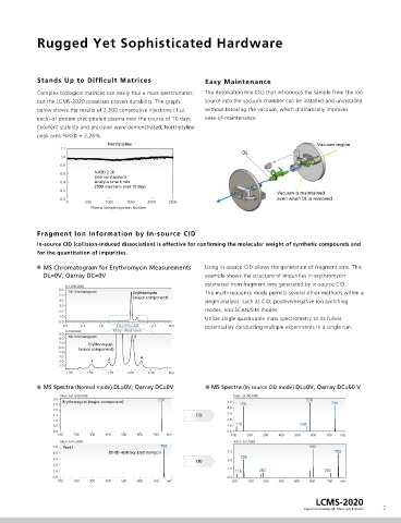
Rapid elution and sharp chromatographic peaks require Ultra Complex biological matrices can easily foul a mass spectrometer, The desolvation line (DL) that introduces the sample from the ion
Fast MS acquisition. The three-second wide peaks in this but the LCMS-2020 possesses proven durability. The graph source into the vacuum chamber can be installed and uninstalled
Inten. (×100,000)
chromatogram were acquired on multiple SIM channels with below shows the results of 2,500 consecutive injections (1 µL without breaking the vacuum, which dramatically improves
4.00 Methomyl Thiodicarb
polarity switching to demonstrate the outstanding quantitative each) of protein precipitated plasma over the course of 10 days. ease-of-maintenance.
3.75
performance of the LCMS-2020. Bentazone (−) Excellent stability and precision were demonstrated; Nortriptyline
3.50 Bentazone (+) peak area %RSD = 2.26%.
3.25 Nortriptyline Vacuum region
Triclopyl 1.2
Polarity switching time Polarity switching time Dymuron (+) DL
15 msec 15 msec 3.00
1.0
Positive-ion Negative-ion Positive-ion 2.75 Dymuron (−) 0.8
measurement measurement measurement
15,000 u/sec 15,000 u/sec 15,000 u/sec 2.50 Iprodione (+) 0.6 %RSD 2.26
Internal standard
2.25 Iprodione (−) 0.4 Analysis time 6 min
2500 injections over 10 days
0.2
Sample: Polarity 2.00 Vacuum is maintained
0.0 even when DL is removed
1 Methomyl: m/z 163 (+) 0 500 1000 1500 2000 2500
1.75 Carpropamid (+)
2 Thiodicarb: m/z 355 (+) Plasma Sample Injection Number
1.50 Carpropamid (−)
3 Bentazone: m/z 239 (−)
4 Bentazone: m/z 241 (+) 1.25
5 Triclopyl: m/z 256 (−) 1.00 Fragment Ion Information by In-source CID
6 Dymuron: m/z 269 (+)
0.75 In-source CID (collision-induced dissociation) is effective for confirming the molecular weight of synthetic compounds and
7 Dymuron: m/z 313 (−) for the quantitation of impurities.
0.50
8 Iprodione: m/z 330 (+)
9 Iprodione: m/z 243 (−) 0.25 MS Chromatogram for Erythromycin Measurements Using in-source CID allows the generation of fragment ions. This
10 Carpropamid: m/z 334 (+) 0.00 DL=0V, Qarray DC=0V example shows the structure of impurities in erythromycin
11 Carpropamid: m/z 378 (−) estimated from fragment ions generated by in-source CID.
0.00 0.25 0.50 0.75 min 6.0 (×1,000,000)
TIC chromatogram Erythromycin The multi-sequence mode permits several other methods within a
5.0 (major component)
4.0 single analysis, such as CID, positive/negative ion switching
3.0
2.0 modes, and SCAN/SIM modes.
1.0 Utilize single quadrupole mass spectrometry to its fullest
Examples of ionization in positive and negative modes 0.0 0.0 0.5 1.0 1.5 2.0 2.5 min potential by conducting multiple experiments in a single run.
(×100,000) Magnified view
9.0
MS Spectra of Bentazone MS Spectra of Dymuron MS Spectra of Carpropamid 8.0 TIC chromatogram 3
7.0 Erythromycin
Inten. (×100,000) Inten. (×100,000) Inten. (×10,000) 6.0 (major component)
5.0 7.0 5.0
Negative 239 [M−H] − 3 Positive 269 [M+H] + 6 Positive + 10 4.0 2 4
1.5 4.0 6.0 334 [M+H] 3.0 1
2.0
5.0
3.0 4.0 1.25 1.50 1.75 2.00 2.25 min
1.0
3.0
2.0 MS Spectra (Normal mode) DL=0V, Qarray DC=0V MS Spectra (In-source CID mode) DL=0V, Qarray DC=60 V
0.5 2.0
1.0 Inten. (×1,000,000) Inten. (×100,000)
1.0 3.0 734 576
0.0 0.0 0.0 2.5 Erythromycin (major component) 5.0 158 734
4.0
150.0 200.0 250.0 300.0 350.0 m/z 150.0 200.0 250.0 300.0 350.0 m/z 150.0 200.0 250.0 300.0 350.0 m/z 2.0
1.5 CID 3.0
Inten. (×100,000) Inten. (×100,000) Inten. (×10,000) 1.0 2.0
5.0 0.5 1.0 116 558
Positive 241 [M+H] + 4 1.25 Negative 313 [M+HCOO] − 7 6.0 Negative 11
4.0 378 [M+HCOO] − 0.0 100 200 300 400 500 600 700 m/z 0.0 100 200 300 400 500 600 700 m/z
1.00 5.0
Inten. (×10,000) Inten. (×1,000)
3.0 4.0 750
0.75 5.0 Peak1 592
2.0 0.50 3.0 4.0 85-(8)-Hydroxy-Erythromycin 7.5 158 750
2.0 3.0 CID 5.0
1.0 0.25 2.0
1.0 2.5
1.0 116 282 703
0.0 0.00 0.0 0.0 0.0
150.0 200.0 250.0 300.0 350.0 m/z 150.0 200.0 250.0 300.0 350.0 m/z 150.0 200.0 250.0 300.0 350.0 m/z 100 200 300 400 500 600 700 m/z 100 200 300 400 500 600 700 m/z
LCMS-2020
6 Liquid Chromatograph Mass Spectrometer 7