Page 6 - Kratos AXIS Nova
P. 6

HIGH SPATIAL RESOLUTION XPS IMAGING                                                                                    XPS FROM SELECTED AREAS                                ION SPUTTER DEPTH PROFILING
         As well as impressive spectroscopic performance, the AXIS   MULTI-TECHNIQUE CAPABILITIES                               Selected area spectroscopy is performed by inserting an   The AXIS Nova  can be configured with a
                                                                                                                                                                                                  2
             2
         Nova  incorporates a spherical mirror analyser (SMA) for high   Whilst the AXIS Nova  is primarily designed for high   aperture into the electrostatic lens column, forming a virtual   standard floating column monoatomic
                                                                                   2
         spatial resolution XPS imaging. In parallel imaging mode the   throughput high performance XPS additional analytical   probe at the surface of the sample. Spectra are acquired from   Ar  ion source or the Gas Cluster Ion Source
                                                                                                                                                                                         +
         spatial distribution of the photoelectrons is retained as they are   capabilities can be added without compromising the   defined selected areas down to 15 μm diameter with improved   (GCIS) depending on the type of sample to be
         projected onto the 2-dimensional delay-line detector.  Stigmatic,   performance. An ultraviolet He-discharge lamp can be   performance over the first generation instrument. Multipoint   profiled. With either ion source ESCApe software allows
         parallel images can be collected in a matter of seconds offering   added to allow collection of ultraviolet photoemission   spectra can be acquired from any position within the field of   compucentric rotation during sputtering to minimise ion induced
         shorter acquisition times and higher spatial resolution than   spectra (UPS) for valence band and work function        view using an electrostatic deflection system. A simple mouse   sample roughening during etching.
         sequential rastered beam or stage mapping approaches.     measurements.                                                click is used to define the analysis position from an image                 +
         An attribute of the SMA is that it operates in fixed analyser                                                          deflecting the virtual probe using electrostatic scan plates. This   The standard monatomic Ar  ion source (Minibeam 4) provides
         transmission mode ensuring that the energy resolution of   Ion scattering spectroscopy (ISS) can be configured as      approach removes the need to move the sample and hence   continuously variable beam energies between 4 keV and 50 eV.
         photoelectron images is constant for all kinetic energies. This is   an additional technique for elemental characterisation   uncertainties introduced with repeated sample translation.  The ion column has a bend to suppress energetic neutrals as
         of particular importance for quantitative imaging applications.   of the outermost surface of the sample. Both the                                                            well as the capability to operate in floating mode for high ion
         Parallel images may also be acquired at lower pass energies to   Minibeam 4 and 6 ion sources can be configured                                                               beam current density at low ion energies. Float mode gives the
                                                                                       +
         improve the energy resolution, analogous to spectroscopy mode,   for use with low energy He  ions and the                                                                     advantage of improved interface resolution and fast etch rates
         allowing chemical state imaging where necessary.          analyser polarity reversed to achieve this                                                                          even at low ion acceleration voltage.
                                                                   acquisition mode simply through the
         The low spherical aberration of the electron optics ensures that   ESCApe User interface.                                                                                     The multi-mode GCIS (Minibeam 6) is capable of generating
                                                                                                                                                                                         +
         the image of the surface can be magnified onto the detector                                                                                                                   Ar  clusters consisting of hundreds or even thousands of Ar
                                                                                                                                                                                         n
                                                                                                                                                                                                              +
         with very little distortion. This ensures high spatial resolution                                                                                                             atoms as well as monatomic Ar  ions for depth profiling and
                                                                                                                                                                                         +
         images. Parallel imaging at the highest magnification gives a                                                                                                                 He  for optional ion scattering spectrometry (ISS). The use of
         guaranteed spatial resolution of 3 μm.                                                                                                                                        cluster ions allows the successful depth profiling of ‘soft’ organic
                                                                                                                                                                                       polymers with retention of chemistry. The energy per projectile
                                                                   R1 SPECTRUM
                                                                                                                                                                                       atom, or partition energy, can be as low as a few electron volts.
                                                                                                                                                                                       It has been shown that these cluster ions sputter material from
                                                                                                                                                                                       the near-surface, causing very little sub-surface damage so
                                                                                                                                                                                       that excellent interface resolution can be maintained through
                                                                                                                                                                                                                          +
                                                                                                                                                                                       multilayer depth profiles of several microns. Ar  cluster mode
                                                                                                                                                                                                                         n
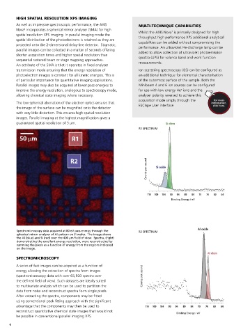
                                                                                                                                Overlaid chemical state images of silicon              depth profiling is also finding application to inorganic depth
                                                                       Counts per second                                        aluminium (red) from a micro-patterned                 cause less preferential sputtering than conventional monatomic
                                                                                                                                dioxide (blue) elemental silicon (green) and
                                                                                                                                                                                       profiling where small (n=500) clusters accelerated to 20 keV
                                                                                                                                device.  The optical microscope image
                                                                                                                                (inset) shows the area analysed using XPS
                                                                                                                                                                                       Ar  ions when etching inorganic materials.
                                                                                                                                                                                         +
                                                                                                                                imaging.  Areas on the XPS image indicate
                                                                                                                                where 15 μm diameter selected area Si 2p
                                                                                                                                core level spectra were acquired.                      Pre-defined ion source operating conditions provided in a look-
                                                                                                                                                                                       up table allow easy use of either ion source. Automation extends
                                                                                                                                                                                       to the argon gas supply for the ion source which can be turned
                                                                                                                                                                                       on and off as required during unattended operation, pressure in
                                                                                   Binding Energy / eV                                                                                 monatomic mode being controlled by an automatically regulated
                                                                                                                                                                                       piezoelectric valve.
                                                                                                                                           ANALYSIS AREA 1


         Spectromicroscopy data acquired at 80 eV pass energy through the   R2 SPECTRUM
         spherical mirror analyser of Al pattern on Si wafer.  The image shows                                                 Counts per second
         the Al (blue) and Si (red) over the 400 μm field of view.  Spectra, (right)
         demonstrating the excellent energy resolution, were reconstructed by
         summing the pixels as a function of energy from the regions indicated
         on the image.

         SPECTROMICROSCOPY                                                                                                                                                          Concentration (%)
         A series of fast images can be acquired as a function of                                                                      Binding Energy
         energy allowing the extraction of spectra from images                                                                                     ANALYSIS AREA 2
         (spectromicroscopy data with over 65,500 spectra over       Counts per second
         the defined field of view). Such datasets are ideally suited
         to multivariate analysis which can be used to partition the
         data from noise and reconstruct spectra form single pixels.
         After extracting the spectra, components may be fitted                                                                                     Counts per second
         using conventional peak fitting approach with the significant
         advantage that the components may then be used to                                                                                                                                                  Etch time (s)
         reconstruct quantitative chemical state images that would not               Binding Energy / eV
                                                                                                                                                                                           +
         be possible in conventional parallel imaging XPS.                                                                                                                            4 kV Ar  depth profile of alternating layers GaAs / Al Ga As, a model single
                                                                                                                                                                                                                        x
                                                                                                                                                                                                                          1-x
                                                                                                                                                            Binding Energy / eV       photon LED device, expanded to show the emitter region corresponding to
                                                                                                                                                                                      layers 15 – 19.
      6                                                                                                                                                                                                                                   7
   1   2   3   4   5   6   7   8