
Page 4 - Solutions for Contaminant Analysis
P. 4
Fourier Transform Infrared Spectrophotometers (FTIR) Raman Spectrophotometers
Every material has a unique infrared absorption spectrum, hence contaminants can be identi ed and characterized by measuring their infrared absorption spectrum Similar to infrared spectra, every material has a unique Raman spectrum that can be used to identify and characterize contaminants by comparing them
with an FTIR system and comparing it against library data. This process can take several seconds to several minutes and is a powerful tool in the analysis of organic against library data.
substances. Raman analysis also requires almost no sample pretreatment. Raman analysis can also use visible light lasers to analyze samples across transparent glass
slides and containers.
˙ FTIR System with Single Reflection ATR Accessory
˙ AIRsight Infrared/Raman Microscope
The single re ection ATR accessory almost entirely removes the need for sample pretreatment
before FTIR analysis. Measurements can be taken while simply holding the object of interest in Raman mode on the AIRsight infrared/Raman microscope can analyze very small
contact with the approx. 1.5-mm diameter prism. contaminants 10 µm and smaller in size not easily analyzed in infrared mode. Raman
Measurements can be collected directly from samples in a variety of forms, including powders, mode can also be used to characterize inorganic materials not easily characterizable in
lms, bulk samples, liquids, and surface deposits. infrared mode (for metal inorganic materials, use the EDX systems mentioned above or
Plastic Analyzer (Uses IRSpirit FTIR Unit)
EMPA systems mentioned below).
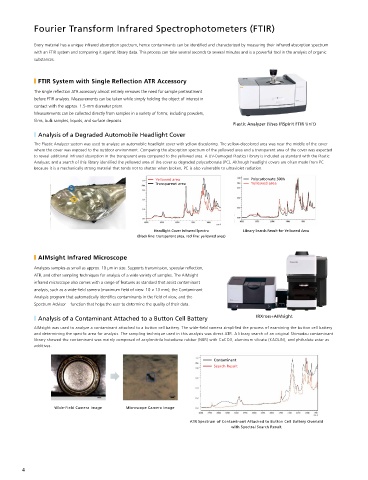
˙ Analysis of a Degraded Automobile Headlight Cover IRTracer-100+AIRsight
The Plastic Analyzer system was used to analyze an automobile headlight cover with yellow discoloring. The yellow-discolored area was near the middle of the cover ˙ Analysis of a Reddish-Brown Contaminant on the Surface of a Medicinal Tablet
where the cover was exposed to the outdoor environment. Comparing the absorption spectrum of the yellowed area and a transparent area of the cover was expected A reddish-brown contaminant was found scattered over an area around 100 µm in size on the surface of a medicinal tablet. Infrared mode with ATR sampling was initially
to reveal additional infrared absorption in the transparent area compared to the yellowed area. A UV-Damaged Plastics Library is included as standard with the Plastic used to analyze the normal area and contaminated area of the tablet, but this approach did not provide useful peaks from the contaminated area. Switching to Raman mode
Analyzer, and a search of this library identi ed the yellowed area of the cover as degraded polycarbonate (PC). Although headlight covers are often made from PC and performing a similar analysis of the normal area and contaminated area of the tablet revealed peaks that seemed to show the contaminant. A spectral search then
because it is a mechanically strong material that tends not to shatter when broken, PC is also vulnerable to ultraviolet radiation.
revealed the adhered contaminant to be iron oxide.
Yellowed area Polycarbonate 500h
Transparent area Yellowed area Normal Area
Contaminated Area
Headlight Cover Infrared Spectra Library Search Result for Yellowed Area
(Black line: transparent area, red line: yellowed area) Raman Spectrum of Contaminant Adhered to Surface of
Medicinal Tablet Overlaid with Raman Spectrum of Normal Area
˙ AIMsight Infrared Microscope
Analyzes samples as small as approx. 10 µm in size. Supports transmission, specular re ection, Electron Probe Microanalyzer
ATR, and other sampling techniques for analysis of a wide variety of samples. The AIMsight
infrared microscope also comes with a range of features as standard that assist contaminant
Irradiates the sample with an electron beam to perform elemental analysis and obtain geometric data on micrometer-order contaminants at high magni cations.
analysis, such as a wide- eld camera (maximum eld of view: 10 × 13 mm), the Contaminant
Electron probe microanalyzers can perform highly accurate and precise elemental analysis across very small and large areas.
Analysis program that automatically identi es contaminants in the eld of view, and the
Spectrum Advisor function that helps the user to determine the quality of their data.
˙ Analysis of a Contaminant Attached to a Button Cell Battery IRXross+AIMsight ˙ EPMA-1720 Series/8050G
AIMsight was used to analyze a contaminant attached to a button cell battery. The wide- eld camera simpli ed the process of examining the button cell battery ˙ Analysis of a Contaminant on a Frozen Pizza
and determining the speci c area for analysis. The sampling technique used in this analysis was direct ATR. A library search of an original Shimadzu contaminant
library showed the contaminant was mainly composed of acrylonitrile butadiene rubber (NBR) with CaCO3, aluminum silicate (KAOLIN), and phthalate ester as A black contaminant attached to a frozen pizza was sampled and subjected to elemental mapping
additives. analysis. Iron (Fe) and chrome (Cr) were detected across a wide area and uorine (F) was detected in
localized areas of the sample. FTIR analysis revealed starch, linseed oil, and uoropolymer material, and
EDX analysis revealed nickel (Ni) in addition to Fe and Cr. The black contaminant is presumed to be burned
Contaminant
vegetable oil mixed with uorine compounds and stainless steel powder originating from the cooking
Search Result
equipment and manufacturing machinery, though this requires veri cation at the manufacturing site. EPMA-1720 Series
Wide-Field Camera Image Microscope Camera Image
ATR Spectrum of Contaminant Attached to Button Cell Battery Overlaid
with Spectral Search Result
EPMA-8050G
4 5