
Page 5 - Pharmaceutical- Guide to Biopharmaceutical
P. 5
Cell Line Optimization
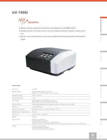
Quantitation of Nucleic Acids UV-1900i
Quantitation of Double-Stranded DNA benefits
- Trace Measurement Using TrayCell and Nano Stick - Cell Line Optimization
click here • Spectra can be acquired at ultra-fast scan speeds up to 29,000 nm/min.
• Sample volume of as low as 0.7 µL can be measured using a TrayCell or Nano Stick
Operating Principle and Features Table 1 Measurement Conditions
Wavelength (Calibration curve): 260 nm, 320 nm cell.
The UV-1900i UV-VIS spectrophotometer features a space-saving and Wavelength range: 220 nm to 330 nm • Nucleic acid concentration can be easily determined using the built-in Biomethod
ergonomic hardware design. The user interface (UI) is displayed on a Scan speed: Low
color touch panel to ensure the system status and operating procedures Sampling pitch: 1.0 nm mode. Culture
can be determined easily with a single glance. The Biomethod mode
includes six types of built-in measurement conditions: 1. Nucleic acid
quantitation, 2. Lowry method, 3. BCA method, 4. CBB (Bradford
method), 5. Biuret method, and 6. UV method. These methods can
be used to measure samples easily for given analytical objectives. The
operation panel screenshot function can be used to easily extract
measurement results without connecting to a computer. A 10 mm
square cell requires a sample volume of approx. 4 mL, but the use of
a TrayCell or Nano Stick cell enables measurement of micro sample Fig. 1 TrayCell
quantities of approx. 2 to 4 μL. Purification
Measurement Method
Double-Stranded DNA Measurement Method Using a TrayCell
Double-stranded DNA was prepared to create 27.5, 55, 110, 220, and
440 ng/μL standard samples (diluted with ultrapure water). Actual 1. Place the sample at the center of
samples were prepared by ethanol precipitation of the same DNA. With the silver area. 2. Install the cap.
the TrayCell, the optical path length can be changed to either 1.0 mm
or 0.2 mm by switching between two types of caps. In this example,
a cap with a 1.0 mm optical path length was used to measure 4 μL of
dripped sample based on the conditions listed in Table 1 (Fig. 1). Characterization
Double-Stranded DNA Measurement Method Using a Nano
3. Place the cuvette in the
Stick Accessory spectrophotometer. 4. Wipe off the sample.
Standard samples and actual samples of double-stranded DNA
Specifications
were prepared using the same method as described for the
TrayCell above. The same measurement conditions were also
Instrument UV-1900i
used, as listed in Table 1. 3 μL sample volumes were measured
Sample volume 10 mm standard cell = 2.5 to 4.0 mL
with the 0.5 mm optical path length of the Nano Stick (Fig. 2). TrayCell = 0.7 to 10 μL, Nano Stick = 2 μL min. Quality Control
Fig. 2 Using a Nano Stick Cell
Wavelength range 190 to 1,100 nm
Results 0.5 Nano Stick-S 1.0 TrayCell
Abs. = 0.0010×Conc. Abs. = 0.0021×Conc. Spectral bandwidth 1 nm
Calibration curves and UV spectral results from measurements using 0.4 R 2 = 0.9999 R 2 = 0.9999 Light source 20 W halogen lamp and deuterium lamp
the TrayCell and Nano Stick are shown in Fig. 3 and Fig. 4. Both Built-in light source auto position adjustment
resulted in calibration curves with high linearity and good measurement Absorbance Absorbance 0.5
accuracy, confirmed by correlation and CV values calculated from 10 0.2 Monochromator LO-RAY-LIGH grade blazed holographic grating in Czerny-Turner mounting
repeated measurements of a 440 ng/μL sample. Detector Silicon photodiode
Sample compartment Internal dimensions: W 110 × D 250 × H 115 mm
Conclusion 0.0 0 100 200 300 400 440 0.0 0 100 200 300 400 440 Pharmacokinetics
Concentration (ng/ L) Fig. 3 Calibration Curve Concentration (ng/ L) Distance between light beams 100 mm
TrayCell and Nano Stick accessories were used with a UV-1900i UV- Dimensions W 450 × D 501 × H 244 mm
VIS spectrophotometer to confirm that micro sample quantities on the 0.50 Nano Stick-S 1.00 TrayCell
order of several microliters can be measured accurately and easily. 0.40 ― 440 ng / µL ― 440 ng / µL Weight 16.6 kg
― 220 ng / µL ― 220 ng / µL
― 110 ng / µL ― 110 ng / µL
― 55 ng / µL 0.50 ― 55 ng / µL Output device USB memory (optional)
― 27.5 ng / µL
― 27.5 ng / µL
Application Examples Absorbance 0.20 Absorbance Extended memory (optional)
Data files saved in text format or UVPC format*
*Files in UVPC format can be read with the UVProbe file viewer, which is a function of LabSolutions UV-Vis, or with UVProbe software. Others
• Evaluating DNA purity based on absorbance ratio
0.00 0.00
• Measuring DNA concentration -0.05 -0.10 Display 24-bit color touch screen
220 250 300 330 220 250 300 330
• Measuring protein concentration Wavelength (nm) Wavelength (nm) Touch pen (standard included)
Fig. 4 Absorption Spectra of Lambda-DNA Touch panel protective sheet (optional)
4 5
index index