Page 5 - Microorganism Species Analysis
P. 5

Microscope                                                                                                                                                                                                           Microscope
                                                                         Morphology
            Microorganism Solutions                                                                                                                                                                                              Microorganism Solutions


                     Microscope Observations of Microorganisms                                                                            Data
                     Cultivation, staining, and microscope observations are widely used for the identification of microorganisms. Generally,
                     observations are performed by optical microscopy combined with staining. However, the detailed morphological features of   Scanning Probe Microscope Observations of Microorganisms
                     bacteria and other micron-order microorganisms are difficult to observe at the resolution level of optical microscopes.
                                                                                                                                           Some scanning probe microscope observations of microorganisms are shown below. The scanning probe microscope   Microorganism Species  Observation of
                     Shimadzu employs a scanning probe microscope to capture more detailed morphological features of bacteria to better meet
                                                                                                                                           offers high-resolution images of microorganisms that cannot be observed by an optical microscope. It requires very
                     the requirements for microorganism observations.
                                                                                                                                           simple pretreatment: dripping the bacteria on to a glass sheet, adsorption of the sample, and air-drying only. It clearly
            Observation of
                                                                                                                                           captures the detailed morphological features of the bacteria shapes and flagella.
          Microorganism Species


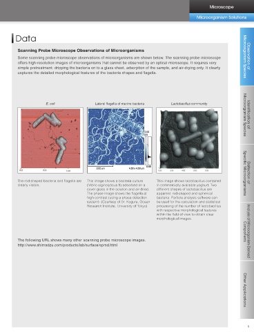
                        Features of the Scanning Probe Microscope                                                                                       E. coli                 Lateral flagella of marine bacteria  Lactobacillus community

                         The scanning probe microscope permits high-magnification observations in air up to several 10,000x magnification                                                                                                              Microorganism Species  Identification of
                         without staining or other pretreatment of the sample surface.
                         The electron optical system with excellent low-acceleration voltage characteristics is suitable for shape observations
                         of the sample surface.
            Identification of
          Microorganism Species


                                                                                                                                           The rod-shaped bacteria and flagella are   This image shows a bacteria culture   This image shows lactobacillus contained   Specific Microorganisms  Detection of
                                                                                                                                           clearly visible.                 (Vibrio alginolyticus 9) adsorbed on a   in commercially available yoghurt. Two
            Detection of
                                                                                                                                                                            cover glass in the solution and air-dried.   different shapes of lactobacillus are
                                                                                                                                                                            The phase image shows the flagella at   apparent: rod-shaped and spherical
          Specific Microorganisms
                                                                                                                                                                            high contrast (using a phase detection   bacteria. Particle analysis software can
                                                                                                                                                                            system). (Courtesy of Dr. Kogure, Ocean   be used for the calculation and statistical
                                                                                                                                                                            Research Institute, University of Tokyo)   processing of the number of lactobacillus
                                                                                                                                                                                                             with respective morphological features
                                                                             Scanning Probe Microscope                                                                                                       within the field of view to obtain clear
                                                                                                                                                                                                             morphological images.


                                                                                                                                           The following URL shows many other scanning probe microscope images.                                        Components  Analysis of Microorganism-Derived
          Components
                                                                                                                                           http://www.shimadzu.com/products/lab/surface/spmd.html


            Analysis of Microorganism-Derived
                                                         Fixing and drying


                                    Bacteria                                         SPM-9600
                                                                                     Scanning Probe Microscope                                                                                                                                         Other Applications


          Other Applications


          4                                                                                                                                                                                                                                              5
   1   2   3   4   5   6   7   8   9   10